How Ultrasound Guidance Reduces Filler Complications
Ultrasound guidance has changed how dermal fillers are placed, making treatments safer and more precise. By allowing providers to see beneath the skin in real time, ultrasound helps map blood vessels, tissue layers, and filler placement. This reduces the risk of complications like vascular occlusion, which can cause serious issues such as skin damage or vision loss. It also ensures fillers are placed in the correct layer for smoother, more balanced results.
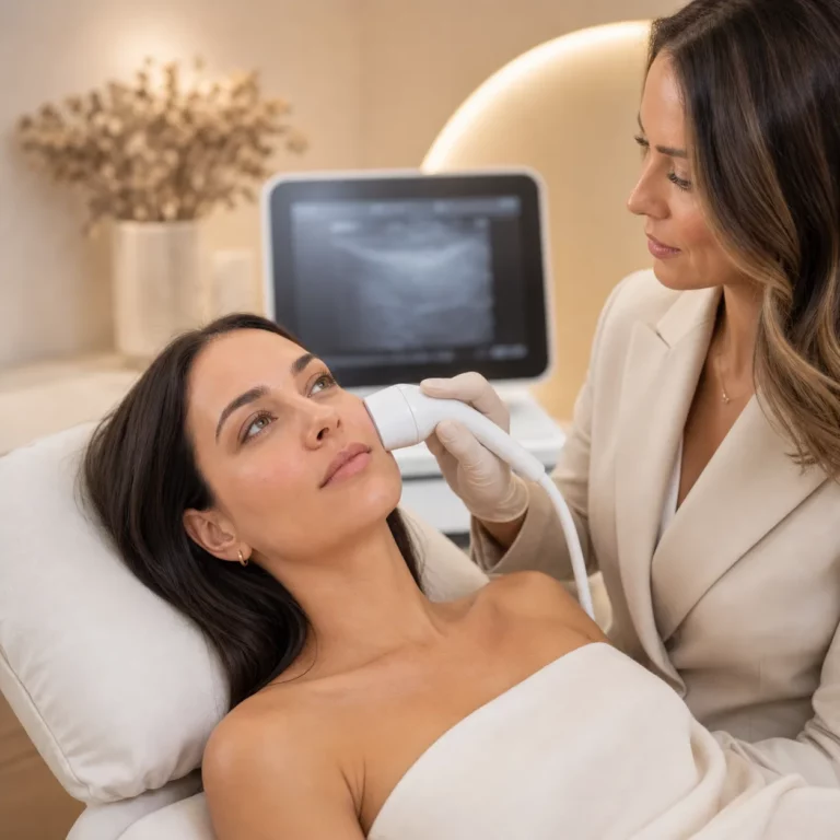
At Calista Aesthetics in Santa Ana, we use ultrasound technology for both filler treatments and corrections. This approach prioritizes safety and precision, especially for high-risk areas like the nose or under-eyes. Whether you’re new to fillers or need to address concerns like lumps or filler migration, ultrasound can make a noticeable difference in your care. Read on to learn how this technique works, why it matters, and what to expect during an ultrasound-guided treatment.
Facial Filler Safety 101: Ultrasound Mapping for Accurate Aesthetic Injections
Common Filler Complications and Their Risks
Dermal filler complications can range from mild cosmetic concerns to severe medical emergencies. One of the most serious risks is vascular occlusion, which occurs when filler is injected into or near a blood vessel, blocking blood flow. Dr. Rosa Sigrist, a lead researcher at the University of São Paulo, explains:
Vascular occlusion events – where the filler is injected into or too close to blood vessels – can be devastating because they can cause tissue death and facial deformity if not treated [4].
In a study analyzing 100 cases of filler complications, ultrasound scans showed absent blood flow in small perforator vessels in 42% of patients and in major blood vessels in 35% of cases [5].
Other complications include nodules and granulomas, which are lumps or masses that may form weeks, months, or even years after treatment. These inflammatory reactions can lead to chronic swelling, tissue hardening, and scarring [6]. Additionally, filler migration and overcorrection can result in visible cosmetic issues like lumpiness, uneven features, or an unnatural look as the filler shifts from its intended placement [6].
What Can Go Wrong with Filler Injections?
Certain areas of the face carry higher risks. For example, injections in the nasal region can lead to severe outcomes like stroke or permanent vision loss if blood vessels are damaged [4]. Without prompt treatment, vascular occlusion can cause tissue necrosis, leading to skin loss and permanent facial deformities [4][5].
Complications can also differ based on timing. Early issues, which appear within days to weeks, may include infections, allergic reactions, or skin necrosis. Late complications, which can arise as long as 21 years after injection, include filler migration, chronic inflammation, and granuloma formation [6]. Nora Nugent, president of the British Association of Aesthetic Plastic Surgeons, emphasizes the importance of preparation:
Mapping out the location of blood vessels undoubtedly provides valuable information ahead of treatment [4].
These risks underscore the importance of advanced imaging techniques and precise planning for safe and effective filler procedures.
How Ultrasound Guidance Reduces Filler Complications
Ultrasound imaging gives providers a real-time view beneath the skin, showing blood vessels, tissue layers, and the position of the needle or cannula during filler injections. This transforms what used to be a blind procedure into a much more precise process.
By mapping each patient’s unique vascular structure, ultrasound helps prevent complications. A study published in Aesthetic Plastic Surgery in October 2025 by Samskin Plastic Surgery Clinic in Seoul revealed that ultrasound imaging can detect variations in the facial artery’s path that aren’t visible to the naked eye or predictable based on surface anatomy [1]. This ability to see beneath the surface is vital for avoiding intravascular injections, which are the main cause of vascular occlusion and tissue damage.
Dr. Rosa Maria Silveira Sigrist, who studied 100 patients with filler-related vascular issues across six international centers, highlighted the impact of ultrasound on precision:
If injectors are not guided by ultrasound, they treat based on where the clinical findings are and inject blindly. But if we can see the ultrasound finding, we can target the exact place where the occlusion occurs [5].
This level of accuracy builds on existing evidence that ultrasound is a key tool for preventing vascular complications.
Ultrasound also ensures the filler is placed exactly where it’s needed. It allows providers to see the soft-tissue layers clearly, ensuring the product is injected at the right depth for the best results. After the injection, a quick ultrasound scan can confirm that the filler is evenly distributed and hasn’t compressed any nearby blood vessels. This precision highlights the difference between traditional and ultrasound-guided techniques.
Standard Injections vs. Ultrasound-Guided Injections
Ultrasound guidance offers a clear advantage over traditional methods. Standard filler injections rely on the provider’s understanding of general facial anatomy, the feel of the needle, and visible surface landmarks. While experienced injectors often develop a strong sense of placement, this approach can’t account for the natural anatomical differences between patients. Even aspiration tests, where the syringe plunger is pulled back to check for blood, can sometimes fail to detect vessel punctures.
In contrast, ultrasound-guided injections provide a visual confirmation of depth and placement. A 2025 study involving 45 Korean patients receiving nasolabial fold correction showed the difference. Twenty patients with volume-deficient folds were treated using ultrasound-guided filler placement with biphasic hyaluronic acid (Lorient No 6 and 4). At the 12-week follow-up, these patients saw their Global Aesthetic Improvement Scale scores improve from “no change” to “much improved”, with no cases of vascular compromise or filler misplacement reported [1].
The distinction lies in visualization versus estimation. Traditional injections rely on the injector’s tactile feedback and experience, while ultrasound provides a live, visual guide. When complications arise, ultrasound guidance allows for targeted, low-dose hyaluronidase to address blockages directly, minimizing tissue damage and improving outcomes compared to broader, less precise treatments.
Using Ultrasound for Filler Dissolving and Complication Treatment
When filler needs to be dissolved – whether due to migration, nodules, or overfilling – ultrasound guidance allows for a much more targeted approach. Instead of injecting hyaluronidase (such as Hylenex) broadly into the area, ultrasound helps providers see exactly where the filler is located. This means the enzyme can be delivered directly to the unwanted deposit, reducing the amount of product needed and minimizing the risk of dissolving filler in surrounding areas. This level of precision is especially helpful when fine-tuning results.
As Dr. Sara Dickie explains:
If a patient wants to dissolve just a little of the filler, that is often very hard because the hyaluronidase spreads into the tissues where it is injected and can dissolve any filler that is there. So, it is hard to be very specific with dissolving. I tell patients that they should consider dissolving filler as all or nothing [7].
Ultrasound imaging not only identifies the exact location of the filler but also confirms its depth and the layer it occupies – whether it’s in the superficial or deep fat. This ensures the enzyme acts only on the target area, which is especially important for correcting subtle asymmetries or achieving partial adjustments rather than removing all filler.
For vascular complications, ultrasound becomes even more critical. It allows providers to locate the exact site of a blockage, ensuring that a precise, lower dose of hyaluronidase can restore blood flow. Research from the Samskin Plastic Surgery Clinic revealed that 38% of patients have variations in their facial artery anatomy, emphasizing the importance of anatomy-based protocols [1]. Ultrasound not only helps avoid complications during filler placement but also provides a reliable tool for addressing them when they arise.
At Calista Aesthetics in Santa Ana’s South Coast Metro, we integrate ultrasound-guided filler dissolving as part of our careful, anatomy-focused approach. Our goal is to resolve concerns with minimal disruption to surrounding tissue while preserving your natural balance and appearance.
Research on Ultrasound-Guided Filler Procedures
Recent studies continue to highlight the role of ultrasound in improving the safety and precision of filler treatments. By preventing vascular complications, ensuring accurate filler placement, and delivering consistent aesthetic results, ultrasound technology is proving to be an important tool in advanced aesthetic procedures.
A study published in January 2026 by Gi-Woong Hong and Kyu-Ho Yi from Samskin Plastic Surgery Clinic in Seoul, Korea, explored its benefits in-depth. The researchers examined 45 patients undergoing nasolabial fold correction using high-resolution ultrasound. They used the technology to map individual variations in facial artery anatomy and applied a precise layering technique with biphasic hyaluronic acid filler. The results? No cases of vascular compromise or filler misplacement. Patients also showed a marked improvement on the Global Aesthetic Improvement Scale (GAIS), progressing from “no change” at the start to “much improved” after 12 weeks [1].
The researchers emphasized:
Ultrasound-guided, anatomy-based filler injection offers improved safety, accuracy in targeting, and continuous monitoring [1].
This approach aligns with Calista Aesthetics’ commitment to patient safety and personalized care.
Ultrasound also plays a key role in treatment planning. It allows providers to distinguish between volume loss, tissue laxity, and muscle-related changes, ensuring fillers are used only where true volume loss exists [1]. This precision helps avoid over-treatment and creates results tailored to each patient’s unique anatomy.
In addition to planning and placement, ultrasound is invaluable for managing complications. It enables early detection of vascular issues and guides the precise administration of hyaluronidase to restore blood flow when necessary [2][3]. As Cameron P. Christiansen and Elie M. Ferneini explain:
New technology and techniques have been discovered to help aid in reducing the risk, including ultrasound for visualization of important anatomical structures [3].
Ultrasound Guidance at Calista Aesthetics
At Calista Aesthetics, located in Santa Ana’s South Coast Metro area, we incorporate advanced ultrasound technology into both filler placement and dissolving procedures. This allows us to take a precise, anatomy-focused approach to every treatment. With high-resolution imaging, we can see soft-tissue details, filler placement, and vascular structures in real time – helping ensure safer, more accurate results tailored to your unique anatomy.
Before administering fillers, we use ultrasound to map the individual pathways of facial arteries. Studies have shown that vascular anatomy can vary significantly from person to person[1]. By identifying these variations, we can avoid high-risk areas and reduce the chances of complications like intravascular injection. Ultrasound also confirms the exact depth of each injection, adding another layer of precision. This same technology is especially useful during corrective filler procedures, where accuracy is key.
Ultrasound also plays an important role in treatment planning. It helps us evaluate whether your concern stems from volume loss, tissue laxity, or muscle-related issues. This detailed assessment allows us to determine whether fillers alone are the right solution or if additional treatments, such as wrinkle relaxers or other options, might be needed[1]. This approach reflects Calista’s commitment to recommending only what makes sense for your goals and anatomy.
Ultrasound guidance is available as a $250 add-on. This technique allows us to precisely locate existing filler and inject hyaluronidase into the correct tissue plane, rather than relying on surface landmarks alone[1]. This targeted approach enhances the accuracy of dissolving procedures, providing better outcomes for corrective treatments.
Whether you’re coming from Costa Mesa, Irvine, Newport Beach, or anywhere in Orange County, our ultrasound-guided methods reflect Calista’s dedication to safe, evidence-based care that prioritizes your natural beauty and well-being.
When to Schedule an Ultrasound-Guided Filler Consultation
Consider scheduling an ultrasound-guided consultation for treatments in areas with intricate vascular anatomy, such as the nasolabial folds, temples, or under-eyes. This step allows for detailed mapping of your unique anatomy, helping to ensure a safer and more customized treatment plan.
If you’ve had fillers previously and noticed issues like lumps, asymmetry, or migration, an ultrasound consultation can be especially helpful. Ultrasound technology allows us to pinpoint the exact location of existing filler, which is crucial for addressing these concerns. Additionally, if you experience symptoms like skin blanching, severe pain, or vision changes, contact us immediately. These signs could indicate vascular compromise, which requires urgent care for your safety [2].
Ultrasound also helps determine whether filler is the best option for you or if another approach might be more suitable. This diagnostic precision ensures that the treatment plan aligns with your anatomy and facial balancing and aesthetic goals.
At Calista Aesthetics, we use ultrasound guidance for both new filler applications and corrections, including our ultrasound-guided Hylenex dissolving service. This reflects our commitment to anatomy-focused care. If you’re in Santa Ana, Costa Mesa, Irvine, Newport Beach, or anywhere in Orange County, visit calistamedspa.com to book your consultation and discover the benefits of precise, personalized treatment.
FAQs
Ultrasound-guided filler injections are not usually more painful than traditional methods. In fact, they can often make the process more comfortable. By allowing for precise placement of the filler, this technique helps to minimize tissue trauma, which may lead to a smoother and more pleasant experience for many patients.
Ultrasound guidance plays a key role when precise visualization of soft tissues, blood vessels, and filler placement is required. It’s particularly helpful in complex or higher-risk areas, where accuracy is essential for achieving safe and effective outcomes while minimizing potential risks.
Ultrasound offers a helpful way to locate and assess existing filler before dissolving it. This can make treatment planning more precise and enhance safety. However, its effectiveness in detecting all older filler deposits can depend on factors such as the type of filler used and where it was placed.